Cool Jobs: Museum science
Samples collected long ago may hold answers to important questions in science and medicine today
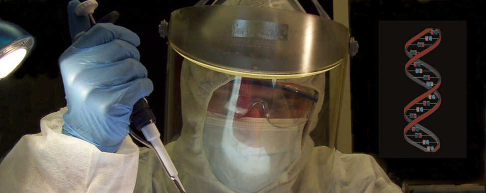
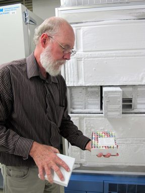
Royal Ontario Museum scientist Oliver Haddrath must wear protective clothing when working with ancient DNA. This ensures his DNA doesn’t get mixed up with the genetic material he is analyzing.
Royal Ontario Museum
When deadly virus outbreaks occur, scientists want to know where the disease is coming from and how to stop it. In their search for answers, some will pay a visit to their local museum. They are not trying to take their minds off the outbreak. Instead, they come to sift through the museum’s historic collections, looking for clues that might help them save lives.
For instance, in the 1990s, there was an outbreak of hantavirus in New Mexico and nearby states. The sometimes-deadly disease causes flulike symptoms and difficulty breathing. At the time, no one knew the source of the outbreak. Some people even suspected terrorists might have released the germs as a biological weapon.
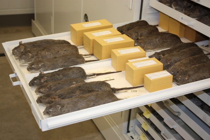
But Robert Baker and his coworkers wondered if a rodent might be to blame. This biologist is a director at the Natural Science Research Laboratory at the Museum of Texas Tech University in Lubbock. Baker knew mice and rats can pass viruses to humans. So he turned to the lab’s stores of dried and frozen tissues for help. Those samples included some collected decades earlier from New Mexico rodents.
His team analyzed deer-mouse lung samples that had been stored in a freezer since the 1980s. Some indeed harbored hantavirus. This showed the germ existed in New Mexico long before the state’s human outbreak developed. The finding suggested biological weaponry was not the outbreak’s source. Most importantly, it pointed to how people could limit infection with the deadly virus: Keep deer mice out of their garages and homes.
Robert Bradley now works as the museum’s curator of mammals. He says the episode taught him an important lesson: Collections like the one he manages let scientists travel back in time to answer important questions. “One hundred years from now, who knows the questions that will be asked? But, he notes, if samples from the past are available, they can help future scientists answer their questions.
Bradley is not the only researcher looking to museum specimens for solutions to new questions in science. Here we get to know Bradley better and meet two other teams using old samples to solve science-based puzzles.
Where diseases come from
Bradley sometimes works with Charles Fulhorst at the University of Texas Medical Branch at Galveston. Fulhorst is a specialist in viral diseases. Together, the two have uncovered the source of other deadly diseases.
For instance, between 2002 and 2010, they analyzed rodent tissues from museum freezers. They identified seven new types of arenavirus in the preserved samples. Arenavirus germs can cause deadly hemorrhagic (HEM or RAAJ ik) fever. Symptoms of this illness range from high fever, muscle aches, loss of strength, and exhaustion to bleeding — not only under the skin but also in internal organs.
Bradley and Fulhorst found that each type of arenavirus is generally carried by a different species of pack rat.
They discovered the source of each germ the same way Baker and his coworkers found carriers of hantavirus. First they tested the museum’s stored rodent tissues for arenavirus antibodies. Antibodies are proteins that the body’s immune system makes to fight a germ or to trigger another type of immune attack. The presence of antibodies indicates the presence at one time of the corresponding, or triggering, virus — in this case, an arenavirus.
When Bradley and Fulhorst found pack-rat kidneys hosting these antibodies, they chopped up those tissues and put them in a Petri dish, a cell-growth chamber. They also placed bacteria in the dish. The reason they added the bacteria: By themselves, viruses can’t reproduce. They must harness the machinery in living cells — like those bacteria — to make more copies of themselves. Over time, arenaviruses grew in the dishes.
The scientists needed a lot of copies of the viruses to look for patterns in the germs’ genetic material. In this case, the genetic material was a type known as RNA. The initials stand for ribonucleic acid. RNA molecules contain an organism’s genetic information — that is, directions for how to grow and function.
The structure of RNA has a slightly different pattern, or sequence, in each type of virus. By comparing those sequences, Bradley and Fulhorst identifed the seven new types of arenavirus. Medical scientists are now developing drugs that might be used in any future outbreaks of these newly discovered germs.
Genetic sleuths
Some scientists turn to museum collections to solve even older mysteries. Oliver Haddrath of Toronto’s Royal Ontario Museum is one such scientist. He’s an expert in ancient DNA. Recently, he studied DNA from the museum’s collection of old bird bones, some more than 1,000 years old. The ancient samples helped him to piece together where flightless birds came from and how they ended up in places such as Australia, New Zealand and Africa.
For the past 50 years, most biologists have accepted the theory that flightless birds — such as kiwis, emus and ostriches — all descended from one flightless ancestor. This ancestor was assumed to live on the supercontinent Gondwana 100 million years ago. Eventually this continent broke up. Parts of it drifted away to make some of the continents — South America, Africa, Australia, Antarctica, regions of Asia — that we know today.
Haddrath and a museum coworker found that this theory was only partially right. The birds indeed descended from a common ancestor that lived on Gondwana. But that ancestor could likely fly.
To figure this out, Haddrath and Allan Baker, head of the museum’s department of natural history, used ancient DNA. Also known as deoxyribonucleic acid, DNA is a molecule inside nearly every living cell. It contains instructions for how each part of the organism should grow and function. (DNA and RNA have different structures.)
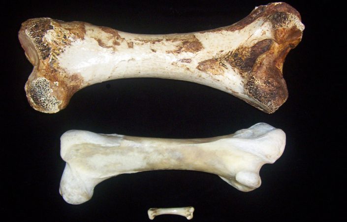
The scientists looked at DNA from a leg bone of a giant flightless bird called a moa. These birds once lived in New Zealand. But people overhunted them, and the species went extinct about 800 years ago.
DNA from the museum’s moa proved quite similar to that of a small bird called a tinamou. Tinamous, which fly, currently live in Central and South America. The genetic similarities between the two birds — one that could fly and another that couldn’t — shows that they are closely related and share a common ancestor.
“It has never been documented that a flightless bird regained flight,” says Haddrath. “It’s not impossible, of course, because they evolved the ability in the first place. But it’s very unlikely.” To evolve means to change gradually over a long period of time.
So, to pass on the ability to fly to the tinamou, its (and the moa’s) ancestor must also have flown, Haddrath and Baker conclude. Over time, moas lost the ability to fly. Tinamous did not. The scientists published their findings in a scientific journal last September.
But what most impresses Haddrath is that the moa is the closest known relative of the small, one-kilogram tinamou. Unlike that tiny flier, moas weighed up to 250 kilograms (550 pounds) and stood 3 to 4 meters (10 to 13 feet) tall.
“Nature never ceases to surprise,” he says.
Identifying crimes
Speaking of surprises, Daniel Antoine of the British Museum in London discovered something unexpected in 2012. A physical anthropologist (like the character Temperance Brennan on the TV show Bones), Antoine is an expert in analyzing human remains.
Using X-rays, he peered into a 5,500-year-old Egyptian corpse that had been naturally mummified. The body, a man’s, has been part of the museum’s collection since 1900. Wrapped in linen and matting, the corpse had been excavated from a shallow grave. Its hot, dry sand had preserved the corpse so well that the skin, bones and internal organs remained relatively intact.
Until now, little else had been known about the man. Then Antoine brought the mummy to a London hospital for a CT scan. CT stands for computed tomography. This scanning technology uses a computer to combine a large series of X-ray images of the body to create detailed 3-D reconstructions. These include images of bones and interior organs.
“The scan took maybe a couple minutes, at maximum,” Antoine recalls. And after glimpsing just a quick picture on the screen, he says, “straightaway I noticed something suspicious on the left shoulder blade.” Shoulder blades are the triangular bones that poke out near the top of your back.
Ultimately, the museum scientist had to wait several weeks for the 3-D computer reconstruction to be completed. Then he could finish a detailed exam.
From his office computer, he looked again and again at the scan to make sure he was interpreting it correctly. He even sent the scan to a researcher in Sweden who uses similar scanning techniques to help solve crimes. That researcher agreed with Antoine — the mummified man had been stabbed in the back. In fact, he probably never even saw his attacker coming.
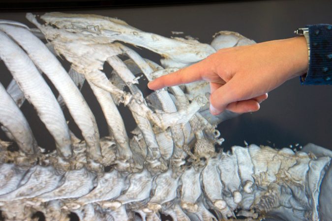
The CT scan showed a cut in the man’s skin over his left shoulder blade. Some sharp, pointed object 1.5 to 2 centimeters (about 0.7 inch) wide had been responsible.
The CT scan also revealed that the stab had shattered a rib just below the victim’s shoulder blade. The blow had been so forceful that it shoved bone fragments into surrounding muscle and injured the man’s left lung and blood vessels.
Because the body showed no signs of healing, the man probably died shortly after being stabbed. The scan turned up no evidence of defensive injuries — signs that the man had fought back. That suggests the victim had no idea — until it was too late — that someone had snuck up on him.
The CT scan also showed where parts of leg or arm bones had fused together. That indicated the victim had only recently finished growing. This placed his age at between 18 and 21 years old. Antoine also saw that the man’s teeth, fully visible for the first time, contained little wear and no dental problems. This further confirmed the victim had been no older than in his early twenties.
Antoine is now eager for scientific advancements that will help him answer more questions he has about the ancient corpse. He wants to scout for ancient viruses or bacteria that would have infected people thousands of years ago.
Like Bradley and Haddrath, he expects museum artifacts like his mummy will eventually give up more of their secrets.
“I want to know how humans adapted and responded to those viruses and bacteria, and the impact population size and climate have on the spread of disease,” says Antoine. “We are only beginning to understand how viruses and bacteria evolve. This is a perfect illustration of the value of these [museum] collections.”
Power Words
antibody Special proteins in the blood that help fight infections, such as those caused by viruses and bacteria.
bacteria A major class of microscopic, single-celled organisms.
curator Someone who manages a collection of items, for instance in a museum, library or art gallery.
DNA Short for deoxyribonucleic (dee OX ee RI boh nu KLAY ik) acid. Genetic instructions inside a living organism’s cells that tell them how to grow and function.
evolve To change gradually over a long period of time.
hemorrhagic Related to major or uncontrolled bleeding, often internally
mummify The process by which a corpse is preserved chemically or through drying. In many cases, communities have intentionally preserved certain members of their society. But bodies of some humans and animals have naturally mummified as the tissues dried out before microbes could degrade them (break them down, as by rotting).
Petri dish A shallow, circular dish used to grow bacteria or other microorganisms.
physical anthropology The type of anthropology, or study of humankind, that deals with human evolutionary biology (how humans have gradually changed over time), physical variation and classification.
viral RNA RNA is short for ribonucleic (RI boh nu KLAY ik) acid. Genetic instructions inside a virus that tell it how to grow and function.
virus A molecule containing genetic information and enclosed in a protein shell. A virus — which can cause illness — can live only in the cells of living creatures.
This is one in a series on careers in science, technology, engineering and mathematics made possible by support from the Northrop Grumman Foundation.